
Hidden Tooth Decay
Images can reveal cavities between teeth, below the gumline or around existing fillings and crowns.
Port Dover Family Dentistry
Digital dental X-rays in Port Dover provide detailed diagnostic images that help identify hidden decay, bone changes and other concerns when clinically needed.
Let’s get you smiling
Tell us how we can help. Our team will follow up to confirm a time.
Digital dental X-rays in Port Dover
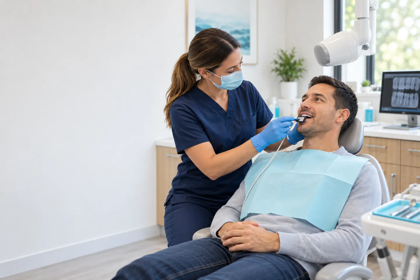
Dental X-rays help your dentist examine areas that cannot be seen directly, including between teeth, beneath existing restorations and around tooth roots and supporting bone. These images can clarify a diagnosis, support treatment planning and help monitor a known condition.
Digital receptors capture detailed images that can be reviewed promptly with you. X-rays are not taken automatically at every visit; the type and number of images are selected after considering your exam, symptoms, history, risk factors and available previous records.
Imaging with a clear purpose
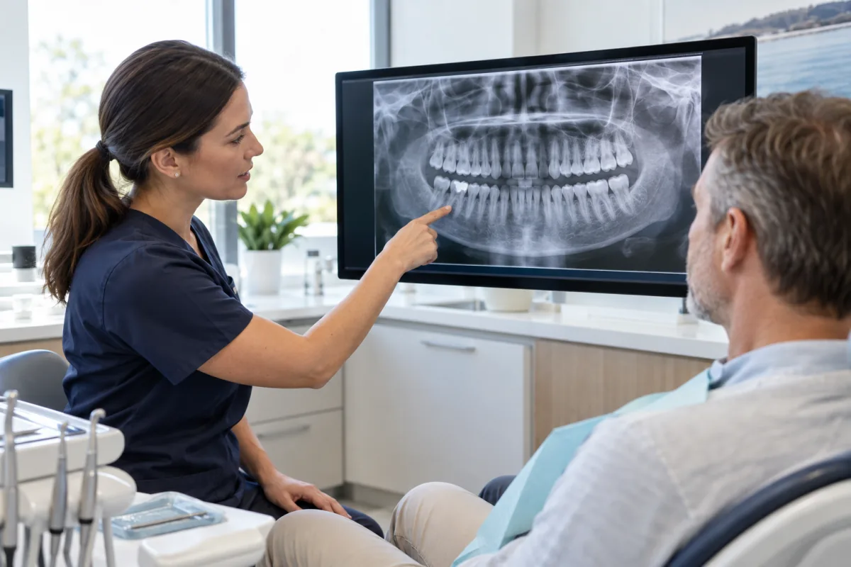
We prescribe dental X-rays when the expected diagnostic benefit justifies the exposure and use the minimum practical number of images needed to answer the clinical question.
Diagnostic detail
Images can reveal cavities between teeth, below the gumline or around existing fillings and crowns.
X-rays can show tooth roots and surrounding areas when investigating pain, infection or a tooth that has been injured.
Bone levels around teeth can be assessed when checking for periodontal changes or planning restorative care.
Imaging may help evaluate unerupted or impacted teeth and the position of teeth in relation to nearby structures.
The right image for the question
Bitewings show the crowns of upper and lower teeth in the same area. They are often used to look between back teeth for decay and to assess the height of supporting bone.
A periapical image shows an entire tooth from crown to root along with nearby bone. It can help investigate pain, trauma, infection or root-related concerns.
A panoramic X-ray provides a broad view of both jaws and all teeth. It may be useful for assessing tooth development, wisdom teeth and other structures that need a wider view.
Purposeful diagnostic imaging
Different X-rays show different structures. The dentist first considers your symptoms, examination findings and available records, then selects the view most likely to provide useful information.
Once the image is captured, we review the relevant findings with you and explain how they affect monitoring, diagnosis or treatment planning.
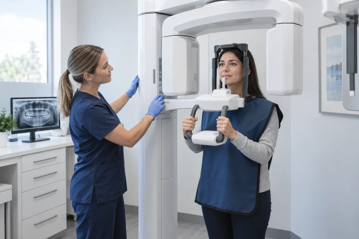
Imaging is recommended to answer a specific question rather than as an automatic part of every visit.
Bitewing, periapical and panoramic images each reveal different areas and levels of detail.
You can see the image and understand why it matters before deciding on next steps.
When imaging can add useful information
An X-ray may be recommended when seeing beneath or between visible surfaces could change the diagnosis, monitoring plan or treatment decision.
Changes between teeth or beneath restorations may not be visible during a clinical examination.
Root and surrounding bone views can help investigate unexplained pain, swelling or a suspected infection.
Images can support evaluation of bone levels when periodontal changes are suspected or monitored.
A wider view may help assess unerupted teeth, wisdom teeth or anatomy relevant to planned care.
Useful recent images from another dental office can often be transferred with your consent and may reduce the need for repeat imaging.
Thoughtful radiation protection
Dental X-rays use ionizing radiation, so each exposure should provide a meaningful diagnostic benefit.
We first consider the clinical exam and any useful recent images. When imaging is needed, equipment settings, positioning and the number of views are chosen to obtain the required information with the lowest practical exposure.
Let us know if you are or may be pregnant, have a strong gag reflex or tender areas, or if another office has taken recent X-rays. We can consider timing, precautions and positioning for your situation.
Helpful information
Dental X-rays use ionizing radiation, so they are prescribed only when the diagnostic benefit is expected to outweigh the small risk. Proper technique and modern equipment help keep exposure to the lowest practical level.
No. The timing depends on your symptoms, examination findings, cavity and gum-disease risk, treatment history and available recent images. Healthy patients may need images less often than someone whose condition is being monitored.
Yes. With your consent, a previous dental office can usually provide relevant images. If they are recent and diagnostically useful, they may reduce the need for repeat imaging.
Tell your dentist if you are or may be pregnant. If imaging is necessary for safe diagnosis or treatment, your dentist will consider the benefits, timing and appropriate radiation-protection measures for your situation.
Intraoral sensors can create brief pressure, especially in a small or sensitive mouth. Tell us if you have a strong gag reflex or tender areas so we can adjust positioning and give you breaks.
Recognized year after year
Best Dentists in Norfolk
Proudly recognized among Norfolk’s top-rated dental practices for six consecutive years.

2021 Winner

2022 Winner

2023 Winner

2024 Winner

2025 Winner

2026 Winner
Stories from our patients
Watch a patient’s experience and browse more five-star reviews from people who have visited our Port Dover dental clinic.
Patient video
A patient shares the welcoming atmosphere and positive experience they found at our Port Dover office.
Google reviews
Swipe or use the arrows
Read all reviewsOur community
Your next visit can start today
New patients and CDCP patients are welcome. Tell us how we can help and our Port Dover dental team will follow up with you.
Evening appointments Monday and Tuesday