
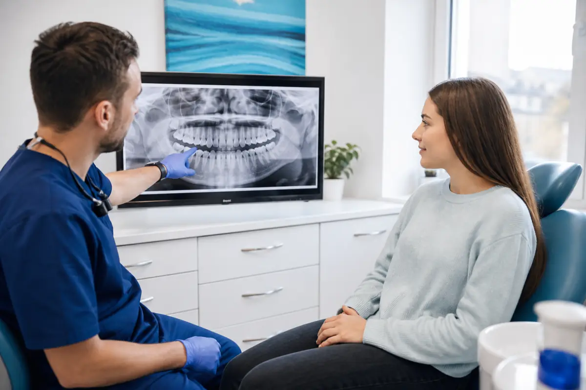
Wisdom teeth removal in Port Dover may be recommended when third molars are impacted, repeatedly infected, decayed or damaging neighbouring structures. It is not an automatic rite of passage. Some wisdom teeth erupt in healthy, functional positions and can be monitored; others remain partly or fully trapped in gum or bone.
The decision considers symptoms, disease, tooth position, cleaning access, root development, nerves, sinuses and overall health. This guide explains evaluation, surgery and recovery so you can participate in an informed decision.
What are wisdom teeth?
Wisdom teeth are third molars at the back of the mouth. They commonly develop and erupt from the late teenage years into the twenties, although timing and number vary. Some people have fewer than four or none.
An impacted tooth does not have enough space or a favourable path to erupt completely. It may be vertical, angled, horizontal or trapped beneath gum and bone. Partial eruption can create a flap that traps plaque and food.
The American Association of Oral and Maxillofacial Surgeons explains that impacted teeth can be symptom-free yet still develop disease, which is why clinical and radiographic monitoring matters.
Signs a wisdom tooth needs assessment
- Pain or pressure behind the last molar
- Swollen, tender or bleeding gum
- Bad taste, drainage or persistent bad breath
- Difficulty opening the mouth
- Recurrent gum infection around a partly erupted tooth
- Decay in the wisdom tooth or second molar
- Food trapping that cannot be controlled
- Cyst or bone change on an x-ray
- Damage to the neighbouring tooth
These symptoms are not specific. Jaw-muscle pain, other molars and throat infections can feel similar. An examination identifies the source.
Does every impacted wisdom tooth need extraction?
No. An asymptomatic impacted tooth without disease may be monitored depending on age, position, future risk and patient preference. “Pain-free” does not prove “disease-free,” but impaction alone does not settle the decision.
Monitoring may include examinations and appropriate x-rays. Removal becomes more compelling with recurrent pericoronitis, non-restorable decay, periodontal damage, cystic change, resorption or other pathology.
Ask the dentist or surgeon to distinguish current disease from predicted future risk and explain the evidence for removal or observation.
How wisdom teeth are evaluated
The clinician reviews symptoms, previous infections, medical conditions, medications, allergies and smoking. Examination assesses eruption, gum pockets, decay, opening and nearby teeth.
A panoramic x-ray often shows tooth angle, roots, bone, nearby second molars, lower-jaw nerves and upper sinuses. A three-dimensional scan may be recommended when anatomy requires more detail, especially when lower roots appear close to the inferior alveolar nerve.
Complex impactions or medical considerations may be referred to an oral and maxillofacial surgeon.
Simple versus surgical extraction
A fully erupted tooth accessible above the gum may be removed with a simple extraction. An impacted tooth often requires a surgical approach: an incision exposes the area, a small amount of bone may be removed and the tooth may be sectioned into pieces. Stitches may be placed.
The AAOMS comparison of simple and surgical extraction notes that position, root shape, surrounding bone and health affect complexity.
Our wisdom teeth extraction service page provides a brief overview.
Anaesthesia and sedation options
Local anaesthetic controls pain at the surgical site. Depending on complexity, anxiety, health and provider authorization, sedation may also be considered. Sedation changes awareness and relaxation; local anaesthetic is still used for pain control.
Ontario has specific requirements for dentist authorization and facility permits at different sedation levels. Discuss fasting, escort, medication and driving restrictions. Never assume you can drive after sedation.
Learn more in our sedation dentistry service overview.
Preparing for removal
- Provide a complete medical and medication history.
- Discuss blood thinners with both prescriber and dental team; do not stop them yourself.
- Follow fasting instructions exactly if sedation is planned.
- Arrange a responsible adult escort when required.
- Fill prescribed medication in advance if instructed.
- Prepare soft foods, gauze and cold packs.
- Avoid scheduling strenuous commitments immediately afterward.
- Ask what to do if you become ill before surgery.
Wear comfortable clothing and avoid jewellery or cosmetics if directed. Confirm which teeth are planned for removal.
The first 24 hours after surgery
A stable blood clot protects the socket and supports healing. Follow individualized written instructions.
Common guidance includes:
- bite on gauze with firm pressure as directed;
- rest with the head elevated;
- use cold packs intermittently outside the face;
- choose cool or lukewarm soft food;
- avoid vigorous rinsing, spitting and straws;
- do not smoke or vape;
- take medication exactly as advised; and
- avoid strenuous activity.
Some oozing is expected. Persistent heavy bleeding despite pressure requires a call.
Eating during recovery
Begin with foods that require little chewing: yogurt, eggs, smooth soup that is not hot, mashed vegetables and soft fish. Avoid hard, sharp, sticky or seedy foods that can enter the socket.
As comfort improves, gradually return to normal texture. Adequate protein and hydration support healing. Do not live only on sugary drinks or ice cream.
Chew away from surgical areas where possible. Numb tissues are easy to bite or burn.
Brushing and rinsing
Clean other teeth carefully while avoiding surgical sites during the earliest period. Begin gentle salt-water or prescribed rinses when directed, often after the first 24 hours. Do not swish forcefully.
Food can collect as lower sockets heal. Your provider may later recommend irrigation, but starting too early can disturb the clot. Follow timing rather than copying another person’s instructions.
Pain, swelling and bruising
Swelling often increases during the first two or three days before improving. Bruising and limited opening can occur. Cold packs are generally used early; later comfort measures depend on instructions.
Use prescribed or recommended pain medication safely. The ADA acute-pain guideline supports non-opioid strategies for many patients, but medication suitability is individual.
Pain that worsens after initial improvement, severe swelling, fever or pus needs assessment.
What is dry socket?
Dry socket occurs when the protective clot breaks down or is lost, exposing bone and nerves. It commonly causes severe throbbing pain a few days after extraction, sometimes radiating toward the ear, along with bad taste or odour.
Risk reduction includes avoiding smoking, straws and vigorous rinsing and following instructions. The AAOMS dry-socket guide advises contacting the surgeon if symptoms develop. Treatment relieves pain and supports healing; it is not fixed by placing aspirin in the socket.
Other risks to understand
Possible complications include infection, prolonged bleeding, damage to neighbouring restorations, sinus communication after upper-tooth removal, jaw stiffness and altered sensation from nerve irritation after lower-tooth surgery.
Numbness of the lip, chin or tongue is usually temporary when it occurs but can rarely persist. Individual risk depends on anatomy. In high-risk lower teeth, a surgeon may discuss coronectomy—removing the crown while leaving roots—when appropriate.
No online statistic replaces your x-ray and health assessment.
When to call after extraction
Contact the treating office for:
- bleeding that does not slow with firm pressure;
- severe pain not controlled as instructed;
- pain that suddenly worsens after several days;
- swelling that continues to increase;
- fever, pus or feeling significantly unwell;
- difficulty swallowing or breathing;
- persistent vomiting or medication reaction;
- numbness that concerns you; or
- fluid passing between mouth and nose.
Breathing or swallowing difficulty is an emergency.
How long does healing take?
Many patients return to school or desk work in several days, while surgical complexity and job demands change timing. Swelling and soreness usually improve over the first week. Gum tissue closes gradually and bone fills the socket over weeks to months.
Feeling better is not the same as complete healing. Continue hygiene and activity precautions for the period advised.
Cost and CDCP coverage in Port Dover
Cost depends on number of teeth, impaction, surgical complexity, imaging, sedation and referral. Ask for an estimate separating extraction and sedation fees.
Current CDCP coverage includes tooth and root removal within program rules. Sedation eligibility and preauthorization vary by level. Co-payments and additional charges may apply.
Wisdom teeth assessment in Port Dover
Port Dover Family Dentistry can assess symptoms and wisdom-tooth position, discuss monitoring versus removal and arrange referral when surgical complexity calls for it.
Call 519-583-2929 or request an appointment. For severe spreading swelling or breathing and swallowing problems, seek emergency medical help.
Frequently asked questions
Can wisdom teeth cause front-tooth crowding?
Crowding is multifactorial, and removal is not a guaranteed way to prevent or correct front-tooth crowding. Orthodontic assessment is appropriate when alignment is the concern.
Can I work the day after surgery?
Some people can after simple removal, while multiple impacted teeth, sedation or physical work require more time. Plan conservatively using your provider’s advice.
Is swelling normal?
Some swelling is expected and often peaks after two or three days. Worsening swelling later, fever, pus, breathing difficulty or severe illness is not routine and needs prompt attention.
About the author
Port Dover Family Dentistry Team
Our team provides family-focused dental care in Port Dover, Ontario. We create practical oral-health education to help patients ask informed questions and feel more confident about care. This article is general information and does not replace an examination or personalized diagnosis.