
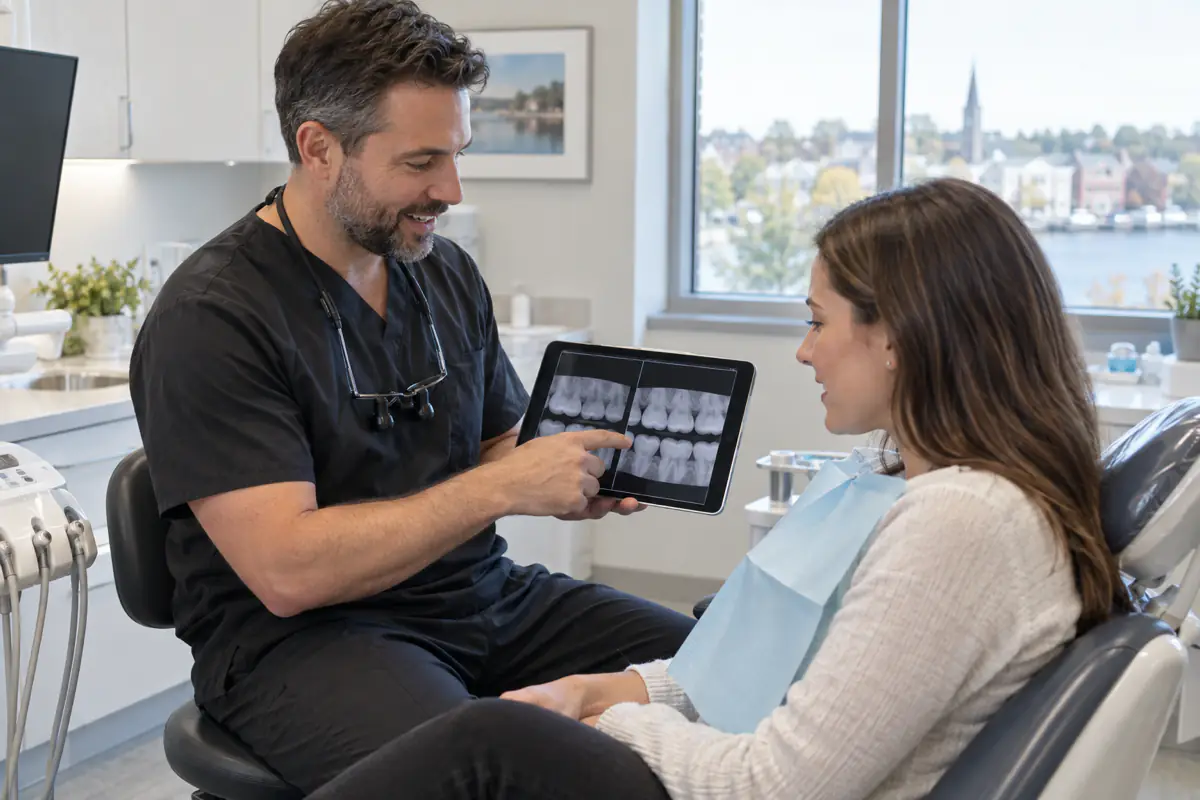
When a dentist recommends dental X-rays in Simcoe, it is reasonable to ask what the image will show and why it is needed now. Radiographs reveal structures that cannot be seen directly during an ordinary examination, including areas between teeth, roots and parts of the jawbone. They can help find disease earlier and guide safer treatment.
X-rays use ionizing radiation, so they should not be taken automatically or merely because a calendar date has arrived. Modern dental imaging is selected according to the patient’s symptoms, age, disease risk, history and planned care. The useful question is not “Are X-rays good or bad?” but “Will this particular image provide information that changes my care?”
What dental X-rays can reveal
Clinical examination shows tooth surfaces, gums, restorations, bite and oral tissues. Some disease remains hidden beneath enamel, under a crown or inside bone. An appropriate radiograph may help identify:
- cavities between teeth or beneath an existing restoration;
- bone changes associated with periodontal disease;
- infection or inflammation around a root;
- an impacted or unerupted tooth;
- root shape before root canal treatment or extraction;
- bone volume and anatomy relevant to implant planning;
- developmental concerns in children and adolescents;
- fractures, resorption or other changes after trauma; and
- cysts, tumours or other unusual jaw findings.
An image is one part of diagnosis. A dark area on a radiograph is not automatically a cavity, and early cracks may not appear. The dentist combines the image with symptoms, examination and tests.
Bitewing X-rays
Bitewings show the crowns of upper and lower back teeth together and part of the supporting bone. The patient closes gently on a holder while the sensor or plate captures the image. These views are particularly useful for detecting decay between teeth and monitoring bone levels.
People with many past cavities, dry mouth, frequent sugar exposure or restoration problems may need bitewings more often than someone with low risk and a long history of health. Children’s intervals also depend on whether contact areas can be seen directly and how quickly the mouth is changing.
Bitewings do not show entire roots. If pain, infection or root anatomy is the question, another type of image may be more useful.
Periapical X-rays
A periapical image usually shows a whole tooth from crown to root tip and some surrounding bone. Dentists use it to investigate a toothache, trauma, root canal concern, deep decay or an unusual finding.
Several images from different angles may be necessary because a three-dimensional tooth is being represented in two dimensions. A second view is not automatically an unnecessary repeat; it may separate structures that overlap in the first image. The reason should still be clear.
Panoramic imaging
A panoramic machine rotates around the head to create a broad two-dimensional view of the jaws, teeth, jaw joints and nearby structures. It can help assess wisdom teeth, development, major pathology and general jaw anatomy.
Panoramic images are not as detailed as bitewings for finding small cavities. A wide view and a close detail view serve different purposes, so one does not necessarily replace the other.
During the scan, positioning matters. Jewellery, glasses and removable appliances may need to come out, and the patient must remain still. A blurred or incorrectly positioned image may be nondiagnostic and require another exposure, which is why careful instruction is part of dose reduction.
Cone-beam CT and three-dimensional imaging
Cone-beam computed tomography, or CBCT, produces a three-dimensional representation. It can be valuable for selected implant, surgical, endodontic, orthodontic and pathology questions. It also exposes a larger volume than many conventional dental images, with dose varying substantially by field size and settings.
CBCT should not be used merely because three-dimensional images look impressive. A smaller, lower-exposure image is preferred when it can answer the clinical question. The American Dental Association’s current radiography guidance recommends incorporating CBCT only when lower-exposure options will not provide the necessary diagnostic information.
If CBCT is suggested, ask what question it will answer, what area will be scanned and how the result may change treatment.
How often are X-rays needed?
There is no universal six-month, annual or multi-year schedule for everyone. A new patient may not need a complete new set if recent diagnostic images can be transferred. Another patient with new pain, rapid decay or advanced gum disease may need targeted imaging sooner.
Selection factors include:
- new symptoms or visible findings;
- cavity history and current risk;
- periodontal condition;
- age and dental development;
- number and age of fillings, crowns or implants;
- dry mouth, diet and fluoride exposure;
- trauma or planned treatment; and
- quality, date and coverage of existing images.
The ADA emphasizes a clinical examination before deciding which images are necessary. Ask the previous dental office to transfer suitable records when changing providers, though new images may still be needed if the old ones are outdated, incomplete or nondiagnostic.
Understanding radiation risk
Ionizing radiation can affect tissue, and exposure from all sources accumulates. Dental imaging contributes a relatively small portion of overall medical imaging exposure, but small is not the same as zero. The ethical approach is to justify each image and optimize the technique.
Radiation risk depends on dose, tissue exposed, age and repeated exposure. Children require particular care because their tissues are developing and they have a longer lifetime ahead. Equipment settings and the image field should be adjusted to the patient and diagnostic task.
Avoid dramatic comparisons that imply radiation is harmless or terrifying. A needed diagnostic image has benefit; an image with no expected clinical value is unnecessary exposure.
How exposure is limited
Dental teams reduce exposure by:
- selecting images from individual need rather than routine habit;
- using digital receptors or other appropriately sensitive systems;
- restricting the beam to the area of interest;
- choosing patient-sized settings;
- positioning the patient and receptor carefully to avoid retakes;
- maintaining and testing equipment; and
- using CBCT only when its additional information is justified.
The traditional term ALARA means keeping exposure “as low as reasonably achievable.” Modern guidance also emphasizes that an image must first be justified and then optimized.
You may notice that shielding practices have changed in some jurisdictions as equipment and evidence have evolved. Current ADA guidance no longer recommends routine thyroid collars or lead aprons for dental imaging because shielding can enter the beam and cause retakes, while beam restriction and technique offer more meaningful protection. Canadian regulatory requirements and office protocols may differ, so ask your dental team to explain the practice being followed rather than assuming a missing apron means safety was ignored.
Dental X-rays during pregnancy
Pregnancy should not prevent diagnosis of urgent dental infection or trauma. Untreated pain and infection can create their own risks. Necessary dental X-rays can generally be obtained with appropriate selection and technique, but the decision remains individualized.
Tell the dental office if you are pregnant or could be. The dentist can determine whether imaging is needed now, whether recent images are sufficient or whether an elective image can reasonably wait. Do not delay urgent assessment of swelling, fever or severe pain because of pregnancy.
If you have questions, the dentist may coordinate with your prenatal clinician. See oral health during pregnancy in Norfolk County for more on gums, morning sickness and safe dental planning.
X-rays for children
Children do not automatically need the same series or interval as adults. Images may be considered when tooth contacts cannot be inspected, decay risk is elevated, eruption is unusual, trauma occurs or orthodontic and developmental questions arise.
The image size, beam and exposure settings should be adapted to the child. A calm explanation and properly sized holder can reduce movement and retakes. If a child cannot tolerate a sensor, the team may consider whether another technique is diagnostically acceptable or whether the image can wait.
Parents can ask what area is being imaged, what the dentist expects to learn and whether an existing image answers the question.
Why an X-ray may not show your pain
Early pulp inflammation, small cracks, muscle pain and some soft-tissue conditions may not appear on a dental radiograph. A normal-looking image does not mean pain is imagined. It narrows the possibilities.
Diagnosis may require cold testing, bite testing, periodontal measurements, magnification, repeat examination or specialist referral. Conversely, an incidental image finding may have no relationship to the symptom. Location and clinical testing help connect evidence to the correct tooth.
Digital enhancement and interpretation
Digital software can adjust brightness, contrast and magnification to help a clinician inspect an image. Enhancement does not create anatomy that was never captured. Overprocessing can make noise look significant, so interpretation relies on training and the original diagnostic data.
Artificial intelligence tools may highlight areas for attention, but they do not replace the dentist’s diagnosis. False positives and missed disease remain possible. Patients should receive an explanation in context, not a treatment recommendation based only on a coloured software box.
Questions you can ask
Before an image, consider asking:
- What type of X-ray is recommended?
- What clinical question will it answer?
- Are recent images available from my previous dentist?
- How will the image affect the treatment decision?
- Is a smaller field or two-dimensional image sufficient?
- What happens if we do not take it now?
These questions support informed discussion. Refusing every image can limit diagnosis, while accepting every image without understanding is not necessary. Shared decision-making means weighing expected benefit and risk.
Reviewing the image together
Ask the dentist to orient the image and show the relevant area. On a bitewing, enamel and fillings appear lighter, while decay may appear darker; bone height can be compared around teeth. On a periapical view, the root tip and surrounding bone are visible.
An explanation should include uncertainty. Very early disease may be monitored, while a clear cavity, bone change or infection may require treatment. A radiograph alone does not determine whether a filling, root canal or extraction is appropriate.
Arrange diagnostic care near Simcoe
Port Dover Family Dentistry serves patients from Simcoe and across Norfolk County. We select dental imaging according to examination findings, symptoms, risk and treatment needs, then review relevant results in plain language.
Call 519-583-2929 or request an appointment if you have tooth pain, questions about transferred records or concerns about recommended X-rays. Tell us about pregnancy, previous imaging and any difficulty tolerating sensors so the visit can be planned appropriately.
About the author
Port Dover Family Dentistry Team
Our team provides family-focused dental care in Port Dover, Ontario. We create practical oral-health education to help patients ask informed questions and feel more confident about care. This article is general information and does not replace an examination or personalized diagnosis.