
Cavities between teeth in Simcoe can develop without a visible hole or obvious pain. Dentists call them interproximal caries. Because neighbouring teeth touch, the early lesion is hidden from a mirror and often from direct clinical view. By the time food begins catching or floss tears, the area may already need treatment.
That does not mean every dark mark on an X-ray is automatically drilled. Very early mineral loss may be managed preventively when the surface remains intact. The decision depends on depth, activity, cavity risk, symptoms and whether the lesion is changing over time.
Why the contact area is vulnerable
Toothbrush bristles clean outer, inner and chewing surfaces well when used correctly. They cannot reliably enter a tight contact. Plaque therefore remains along the side walls and just below the contact point unless floss or another interdental tool disrupts it.
Bacteria in plaque metabolize sugars and starches, producing acids. Repeated acid exposure removes calcium and phosphate from enamel. Saliva and fluoride help replace minerals, but frequent snacking or sipping can keep the balance moving toward loss.
The lesion usually begins beneath the contact and spreads through enamel toward dentin. Two adjacent teeth can be affected by the same plaque-retentive area.
Why you may not feel anything
Enamel has no nerves. Early decay can progress silently. Sensitivity may begin after the lesion reaches dentin, but even then symptoms are inconsistent.
Possible signs include:
- floss repeatedly shredding in one contact;
- food packing between the same teeth;
- sensitivity to sweets or cold;
- a dull ache after eating;
- a dark shadow visible through enamel;
- bad taste near one area; or
- a broken ridge as the unsupported surface collapses.
These signs are not specific. A rough filling, tight contact, gum inflammation or crack can cause similar symptoms.
How bitewing X-rays help
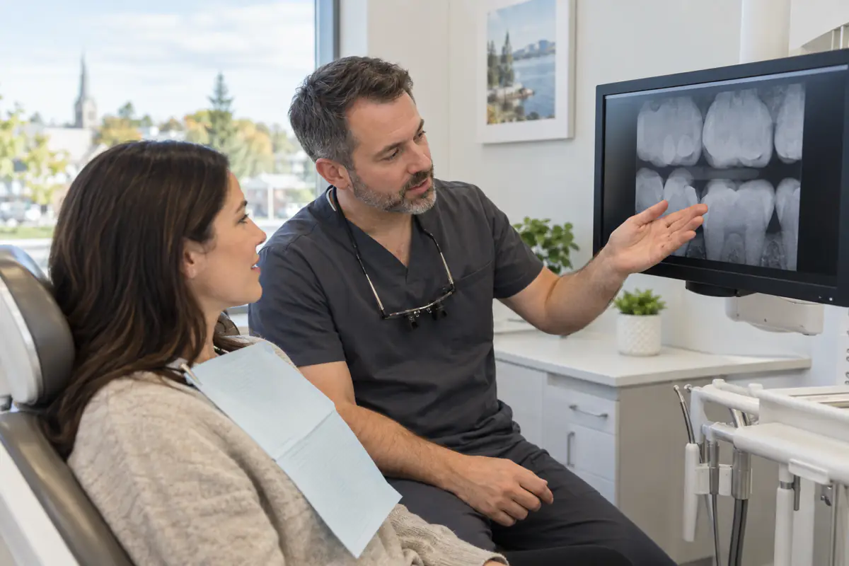
Bitewing radiographs show the crowns of upper and lower teeth and the supporting bone between them. When X-rays pass through an area that has lost mineral, it appears darker than sound enamel. The pattern and depth help the dentist estimate whether decay is limited to enamel or has entered dentin.
Images are prescribed according to risk and clinical need, not automatically at every visit. A person with recent cavities, dry mouth or extensive restorations may need a different interval from someone with consistently low risk.
X-rays can underestimate the true physical size of a cavity and cannot reveal with certainty whether the surface has collapsed. They are interpreted alongside the examination and history.
What early mineral loss looks like
On an X-ray, early interproximal decay often appears as a small triangular dark area in enamel. At this stage, the surface may still be intact. The goal is to stop acid attacks and allow fluoride and saliva to return minerals.
Management may include more effective interdental cleaning, fluoride toothpaste, professional varnish, dietary changes and monitoring with future images. Some practices use resin infiltration or other microinvasive approaches for selected lesions.
The plan depends on activity. An unchanged lesion in a lower-risk patient is different from one that visibly progressed within a short interval.
When a filling is needed
Once the enamel surface cavitates, plaque can live inside a space that home care cannot clean. Decay entering dentin is also more likely to progress. The dentist removes softened, infected structure and restores the tooth with an appropriate material.
To access a cavity between back teeth, the preparation often enters through the chewing surface and extends to the side. The dentist rebuilds the contact so food does not pack and floss can pass with light resistance.
The NIDCR dental-fillings resource explains that fillings repair cavities after decayed tissue is removed. Composite and amalgam are among the available materials; selection depends on the tooth and clinical situation.
Why waiting can mean a larger restoration
An early lesion may need prevention or a small filling. If decay spreads under the enamel, the outer shell can appear intact while dentin weakens. Eventually a ridge breaks, leaving a larger defect.
Deep decay can inflame or infect the pulp. The tooth may then need root canal treatment and a crown rather than a simple filling. If too little sound structure remains, extraction may become necessary.
Pain is a late and unreliable threshold. Treating based on documented progression and structure can preserve more natural tooth than waiting for symptoms.
How dentists judge whether a lesion is active
Activity assessment combines several clues:
- change compared with older X-rays;
- plaque accumulation and gum condition;
- surface appearance where visible;
- recent cavity history;
- fluoride exposure;
- saliva flow;
- diet frequency;
- orthodontic appliances; and
- the person’s ability to clean the contact.
A single image does not tell the entire story. Bringing previous radiographs from another office can help establish whether an area is stable or progressing.
Daily cleaning between teeth
Use floss, an interdental brush, water flosser or another device recommended for the space. Tight contacts usually suit floss. Open spaces, bridges or orthodontic appliances may be easier with a correctly sized interdental brush or threader.
For floss, slide gently through the contact, curve it into a C shape around one tooth and move up and down. Repeat against the neighbouring tooth. Snapping straight into the gum misses tooth surface and can injure tissue.
The NIDCR oral-hygiene guidance recommends regular interdental cleaning because brushing alone does not remove all plaque.
Does a water flosser count?
A water flosser can reduce plaque and inflammation and may help people with braces, bridges or dexterity limitations. It does not remove established tartar or restore a cavity. Technique, pressure and tip placement affect results.
Some people benefit from combining it with floss or an interdental brush. Ask a hygienist to evaluate plaque after your normal routine rather than deciding from how clean the mouth feels.
The best device is one that reaches the surface safely and is used consistently.
Fluoride reaches where brushing cannot
Fluoride in toothpaste mixes with saliva and can support remineralization throughout the mouth, including contact areas. Brush twice daily and follow professional advice about rinsing afterward.
People at higher risk may be offered fluoride varnish, rinse or prescription toothpaste. Concentration and frequency should be individualized, especially for children.
Fluoride strengthens and repairs early mineral loss; it does not fill an established hole. A cavitated lesion still needs mechanical treatment.
Diet frequency matters
Every fermentable-carbohydrate exposure gives plaque bacteria another opportunity to produce acid. Slowly sipping sweet coffee, juice, pop or sports drinks can be more damaging than consuming the same amount with a meal because recovery time is shortened.
Crackers, chips and sticky dried fruit also supply starch or sugar and can remain around contacts. This does not mean they must never be eaten. Limit frequency, choose water between meals and combine snacks with foods that do not extend acid exposure.
Sugar-free gum can stimulate saliva for some people, but it is not suitable for every jaw condition or appliance.
Dry mouth increases interproximal risk
Saliva clears food, buffers acid and supplies minerals. Medicines, dehydration, autoimmune disease and cancer treatment can reduce flow. Decay may then appear at contacts, gumlines and exposed roots even in someone with little previous history.
Report persistent dryness. A dentist or physician can review possible causes and recommend saliva substitutes, additional fluoride or other strategies. Do not stop prescribed medication on your own.
Avoid using sugary candy or acidic lemon drops for moisture. They can worsen the very risk you are trying to manage.
Existing fillings can develop recurrent decay
An interproximal filling has a margin near the gum and contact. Plaque can collect if the margin becomes rough, the contact opens or home cleaning is inconsistent. Decay may form at the edge while the centre of the restoration remains intact.
Floss that suddenly catches, food packing or a new shadow deserves assessment. Not every stained margin is decay, and restorations should not be replaced solely because of age.
Repair may preserve more tooth in selected cases. Replacement is recommended when disease or structural failure makes it necessary.
Braces create additional hiding places
Fixed appliances complicate access under wires and around brackets. A floss threader, orthodontic floss, interdental brush and water flosser can each serve a role. Fluoride toothpaste is essential.
White-spot lesions often begin around brackets on visible surfaces, while cavities can still develop between teeth. Orthodontic visits do not replace examinations and X-rays from the family dentist.
Coordinate care if a cavity is found. The wire may need temporary removal or adjustment so the dentist can restore the contact correctly.
Children and cavities between molars
Children’s back teeth often develop tight contacts after the primary molars touch. A brush cannot clean those sides. Caregivers should begin flossing contacts the child cannot manage.
Primary-tooth cavities matter. They can cause pain and infection, and a broken contact may allow neighbouring teeth to shift. Treatment depends on depth, the child’s age, cooperation and how long the tooth should remain.
Bitewing images are selected based on the child’s contacts and risk, not simply age. A dentist explains when the expected information justifies the image.
Food trapping does not always mean decay
Food can pack because of an open contact, gum recession, tooth movement or restoration shape. Repeated pressure inflames the gum and creates soreness that resembles toothache.
Floss gently after meals and arrange an examination. The solution may be a filling repair, bite adjustment, periodontal care or coaching on a different cleaning tool.
Do not widen the space with toothpicks or metal devices. That can injure the papilla and make trapping worse.
Common misconceptions
“If I cannot see a hole, there is no cavity.” Contact surfaces are hidden, and decay may spread under apparently intact enamel.
“Flossing makes cavities disappear.” Cleaning can help arrest an intact early lesion but cannot rebuild a cavitated surface.
“Every dark X-ray spot needs a filling.” Depth, activity, risk and surface integrity guide the decision.
“No pain means it can wait forever.” Pain often begins after the lesion has become deeper.
“A crown prevents all future decay.” Natural tooth remains at the crown margin and can still decay.
When symptoms are urgent
Call promptly for spontaneous pain, lingering hot or cold sensitivity, pain that wakes you, a broken tooth, gum swelling, fever or a bad taste. Severe facial swelling, difficulty breathing or swallowing, or swelling near the eye or neck requires emergency medical care.
A small asymptomatic lesion is not a hospital emergency, but it still deserves a risk-based plan and monitoring. Keep recommended recall and imaging appointments.
Assessment for cavities near Simcoe
Port Dover Family Dentistry provides examinations and restorative care for patients from Simcoe and surrounding Norfolk County communities. We can review current and previous images, explain whether a contact lesion appears early or cavitated, and discuss preventive and restorative choices.
Call 519-583-2929 or request an appointment. For more detail about restoration recovery, see our dental filling aftercare guide.
Frequently asked questions
Why do I have a cavity if I brush twice a day?
Brushing does not predictably clean tight contacts. Interdental cleaning, fluoride, diet frequency, saliva and individual anatomy all affect risk.
Can a cavity spread from one tooth to the next?
The hole itself does not jump between teeth, but the same plaque and acid environment can create lesions on both surfaces facing one another.
Why does the dentist want to compare old X-rays?
Comparison shows whether mineral loss is stable or progressing. That evidence helps distinguish a lesion suitable for monitoring from one that needs treatment.
About the author
Port Dover Family Dentistry Team
Our team provides family-focused dental care in Port Dover, Ontario. We create practical oral-health education to help patients ask informed questions and feel more confident about care. This article is general information and does not replace an examination or personalized diagnosis.